
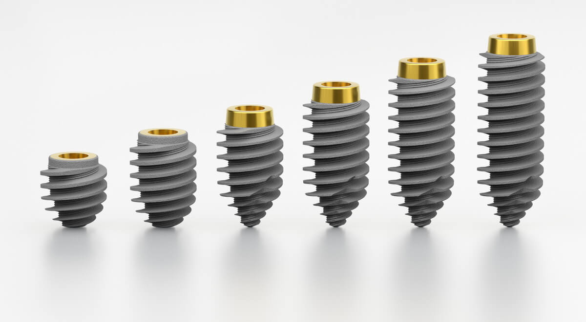
Choosing an implant fit for each situation might be a challenging task. The implant has to provide easy handling, excellent primary stability, and better stability reinsurance in soft bones. Read more and find out more about R55xx implants – a two-piece implant with a self-tapping thread. Measuring 5.5mm in diameter and required length range from 6mm to 16mm for the variety of choice you seek.
Anchorage guarantees stability
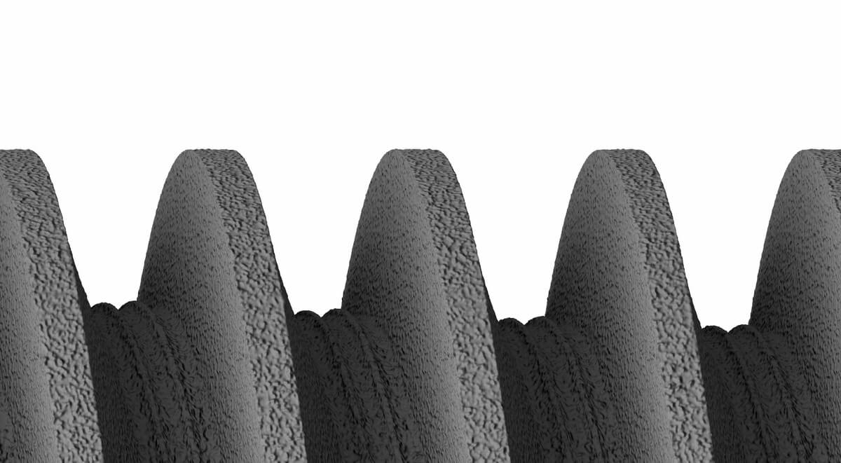
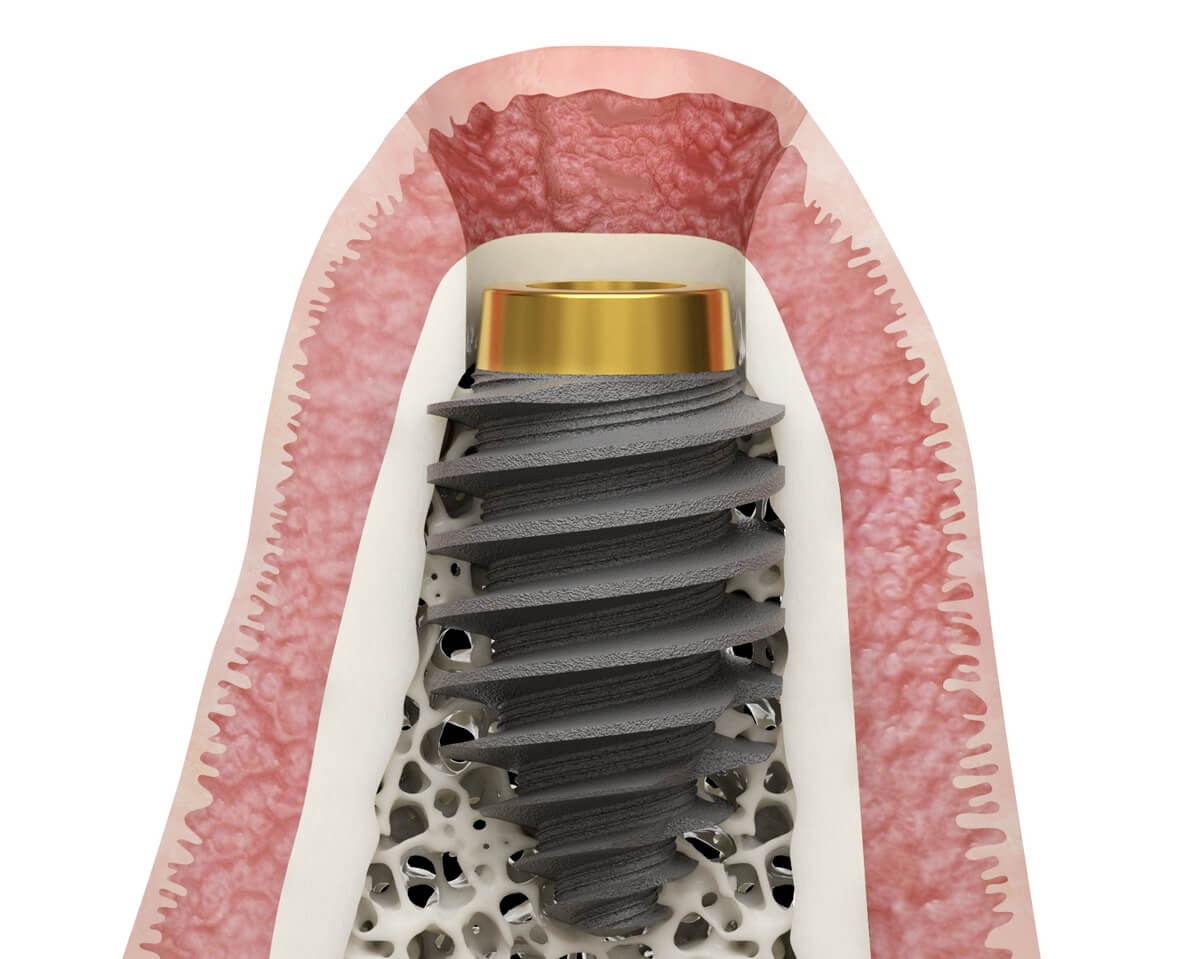
ROOTT R product line is based on excellent primary stability. Thus, the R55xx series is an excellent choice for cases needing firm anchorage. This implant has one step of thread, which provides better stability and more condensation of bone.
Compared to similar diameter implants, R55xx has better initial stability than the rest. The implant is ideal for post-extraction sites, and wide and soft bone types, making it a wise choice for immediate implantation in the molar area. In addition, high strength provides patients comfort and makes specialists work simple.
Ensuring confidence
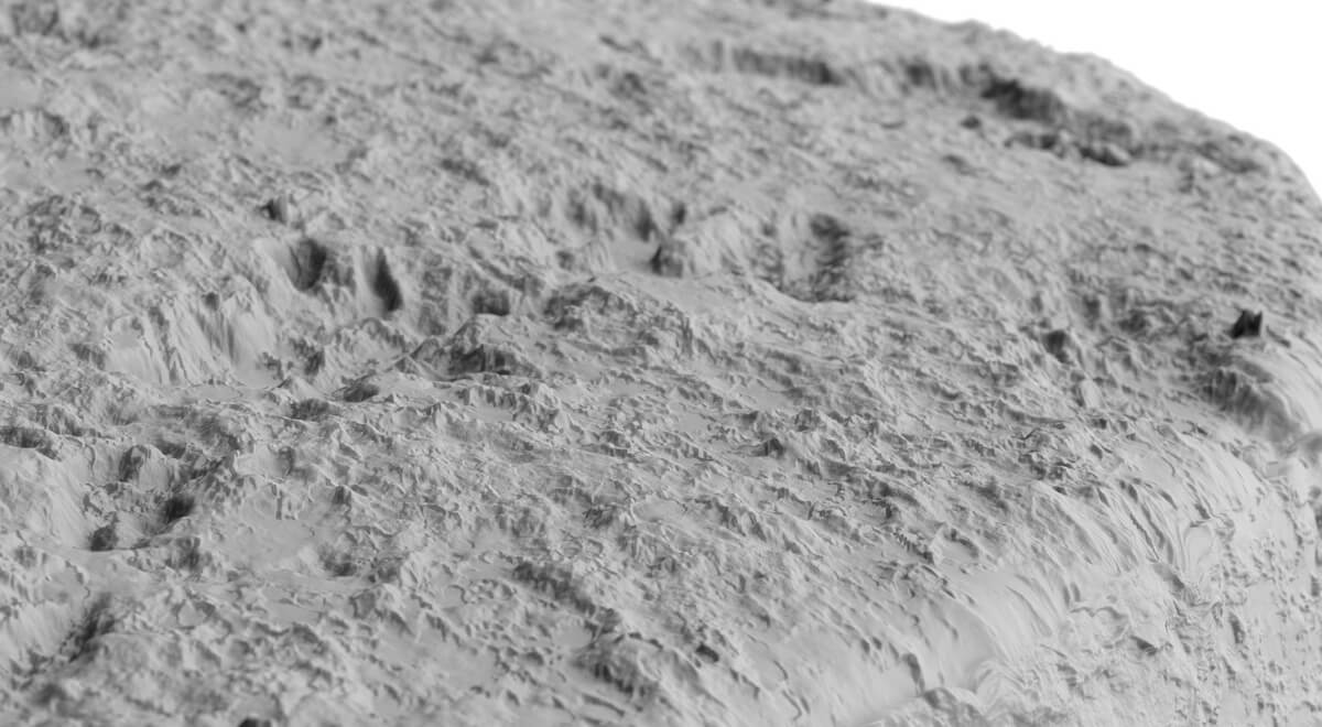
The blasted thread which has a micro-rough surface ensures osseointegration between bone and implant. It is worth mentioning that R5506 and R5508 are fully RBM blasted and R5510 up to R5516 have anodized neck parts.
Important to mention that implants R5506 and R5508 have a one-step thread, and R5510 and R5516 have a two-step thread.

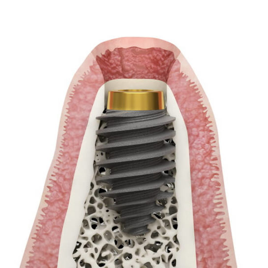
The implant has a back-tapered neck which is good for cortical bone preservation.
Dr. Karampinis Triantafyllos
This implant is in a particular shape resembling a barrel – if put in the socket, its threads quite often start to engage with lateral alveolar cortical bone and achieve better primary stability. The design is anatomical to consider the upper part of the bone, which is most sensitive to infections. Therefore, the implant replicates the width of the bone. With less width at the top and more width in the middle, the implant adapts to the contact of the bone. Also, the lack of micro threads in the anterior part of the implant and decreases the chance of periimplantitis.
Threads for a simple protocol
After some time if gingiva descends, the implant’s neck exposes. In that case, there’s no need to replace the implant – it will be possible to add appropriate abutment accordingly. In addition, an anodized top helps to incorporate the prosthesis lower, increasing the better aesthetic and ensuring mechanical stability consequently.
For doctors that choose immediate implantation due to the anatomy of the implant additional steps of implantation can be avoided which results in a simpler surgical protocol.
Clinical cases
Courtesy of Henri Diederich, Luxembourg
For the 65-year-old patient, we have opted for treatment consisting of 2 pieces of ROOTT R implants in the maxilla in a soft bone.
The unique design of the R5510 with cutting threads gives a strong anchorage in a spongious bone, which was a great option for primary stability.
Here, in this case, I put an additional pterygoid implant with cortical anchorage to have security and long-term result.
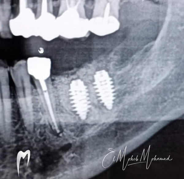
Courtesy of Mohamad El Moheb, France
In this case, we performed extraction and immediate implantation per the patients‘ wishes for comfort. When conducting the protocol, it is easier to implant it in extraction sockets, and the design fits the requirements of the procedure.

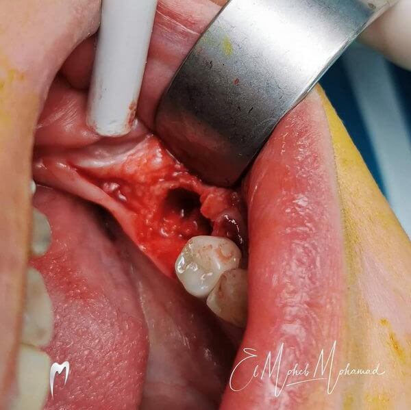
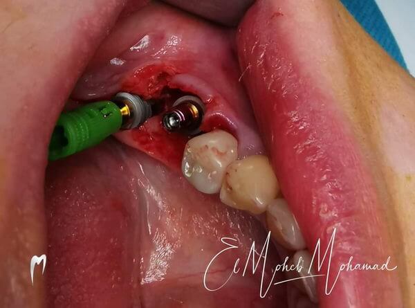
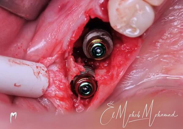
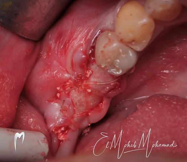
Implantation in the extraction site. Two R5510 were chosen for better primary stability
Courtesy of Davit Manjgaladze, Sakartvelo
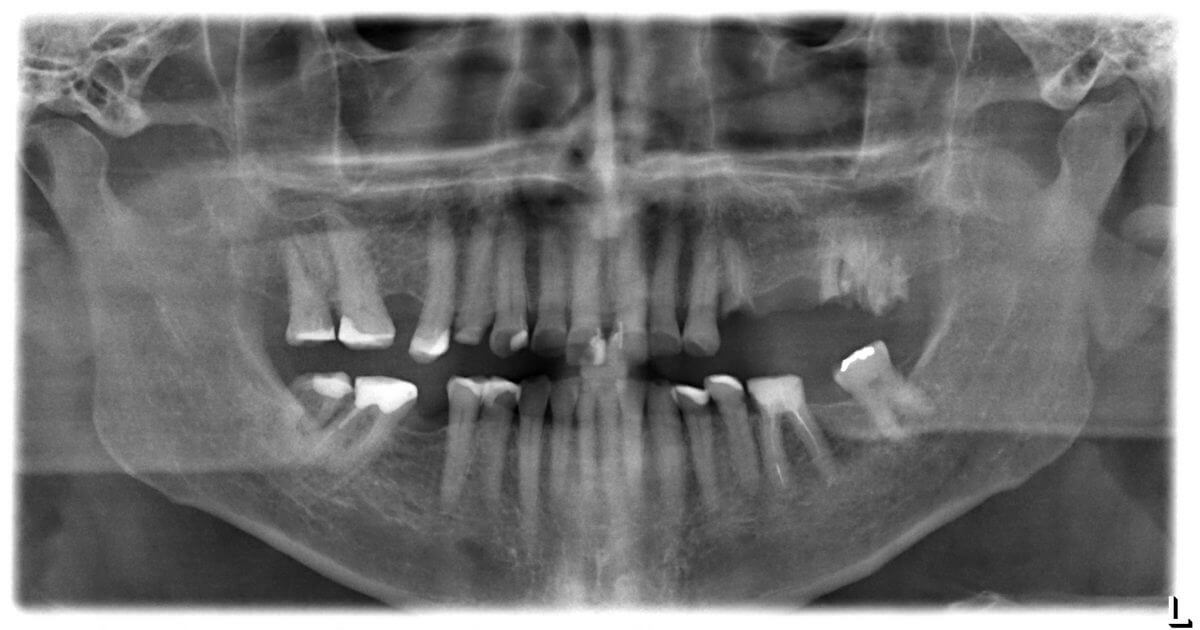
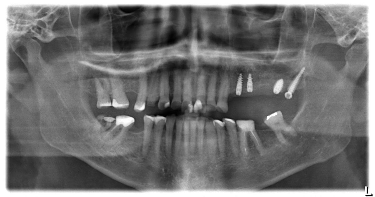
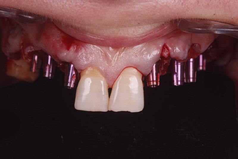
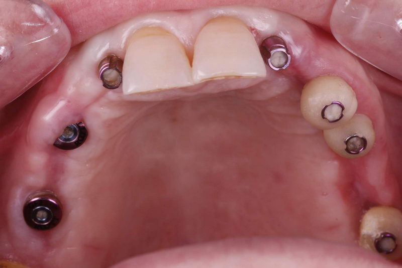
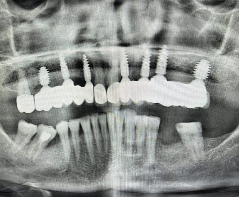
65 years old lady was suffering from esthetic and functional problems caused by loss of teeth. Our proposed and performed treatment options consisted of teeth extraction and implantation with ROOTT system two-piece implants. In addition, we have implemented soft tissue and bone grafting and loaded with zirconia cement-retained bridges after 4 months.
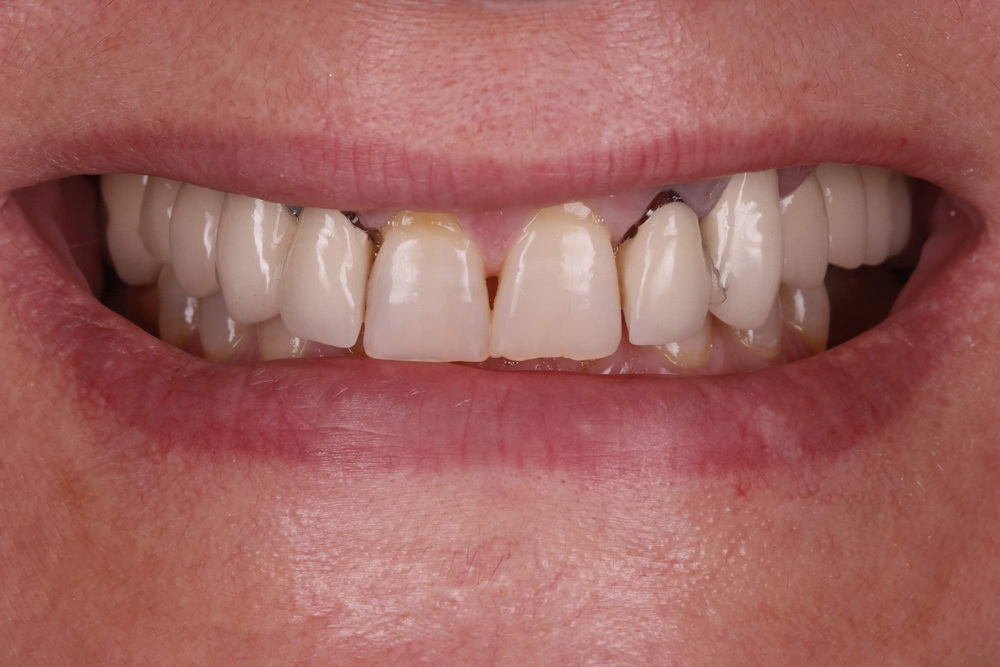
Initial situation
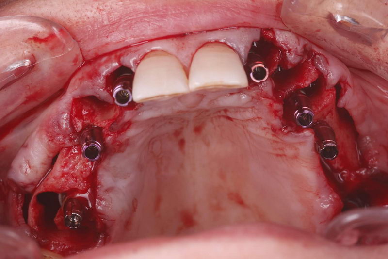
6 implants were placed using the flap method
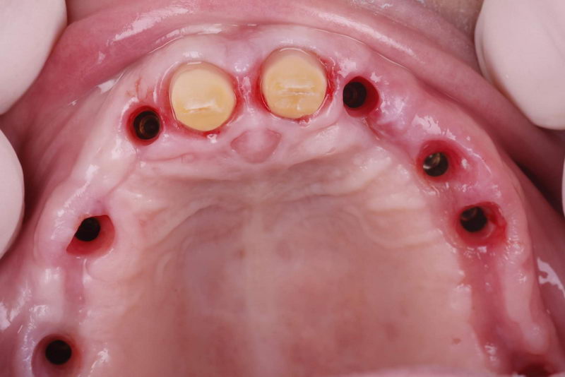
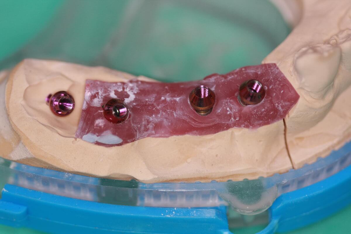
For gingiva formation custom healing abutments with multifunctional part CRE and healing abutments GF2 were used
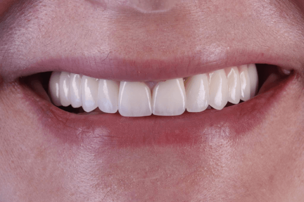
Final restoration
Courtesy of Alexandre Rovisco, Portugal
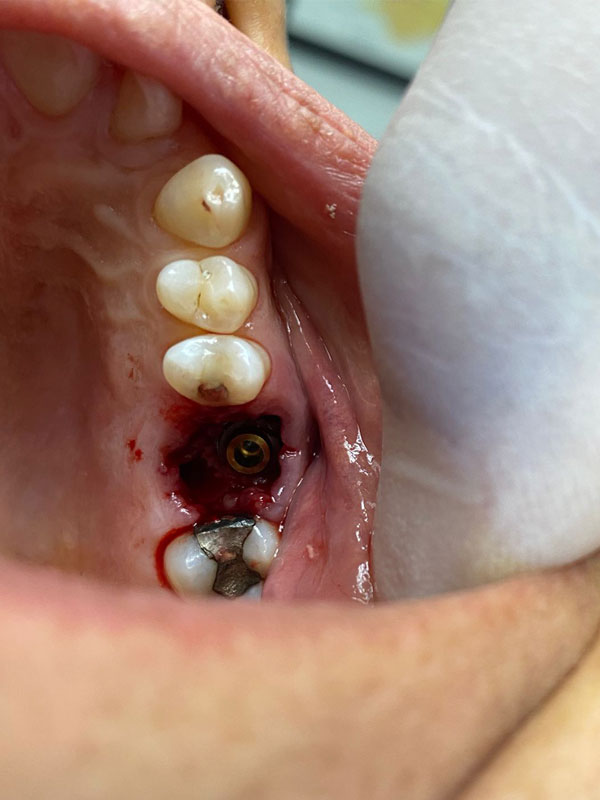
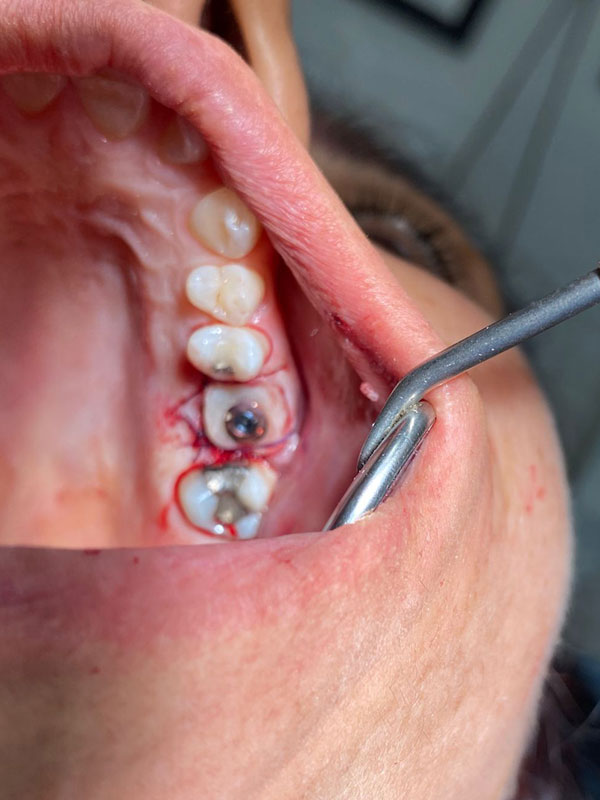
53 years old woman came into clinic with a root fracture was caused by the bad position of a screw. Atraumatic extraction was made to keep the septum. For implantation torque of 70 and implant, R5510 was chosen for the extraction site because of the wide threads which ensure good primary stability. The provisional was made with the Cervico system.

Courtesy of Dainius Karpavičius, Lithuania
53 years old woman came into clinic with a root fracture was caused by the bad position of a screw. Atraumatic extraction was made to keep the septum. For implantation torque of 70 and implant, R5510 was chosen for the extraction site because of the wide threads which ensure good primary stability. The provisional was made with the Cervico system.
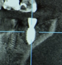
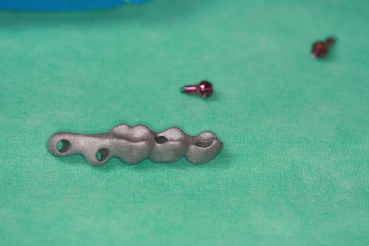
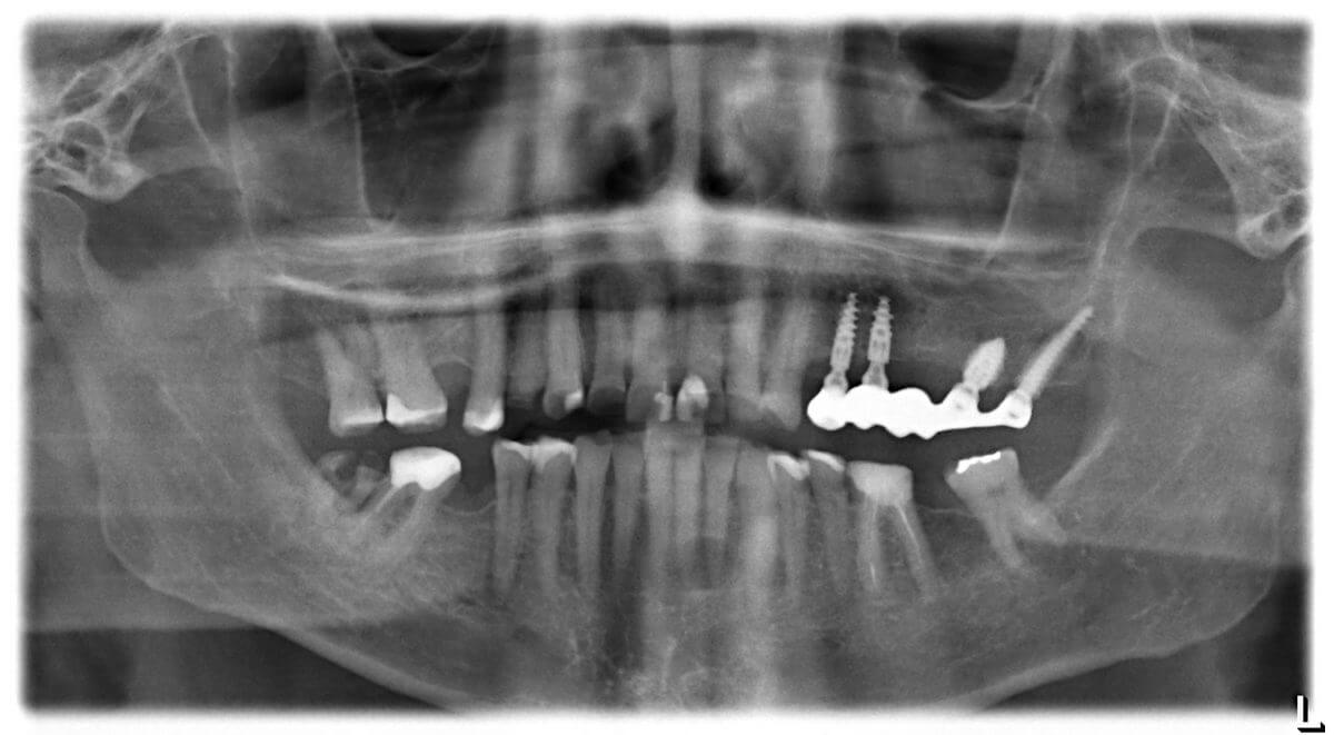
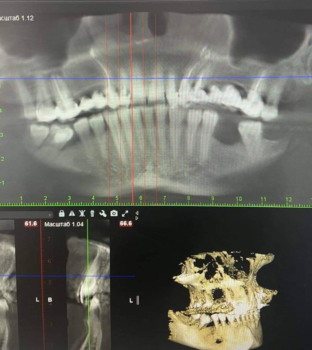
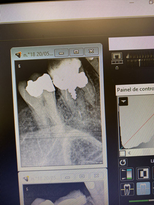
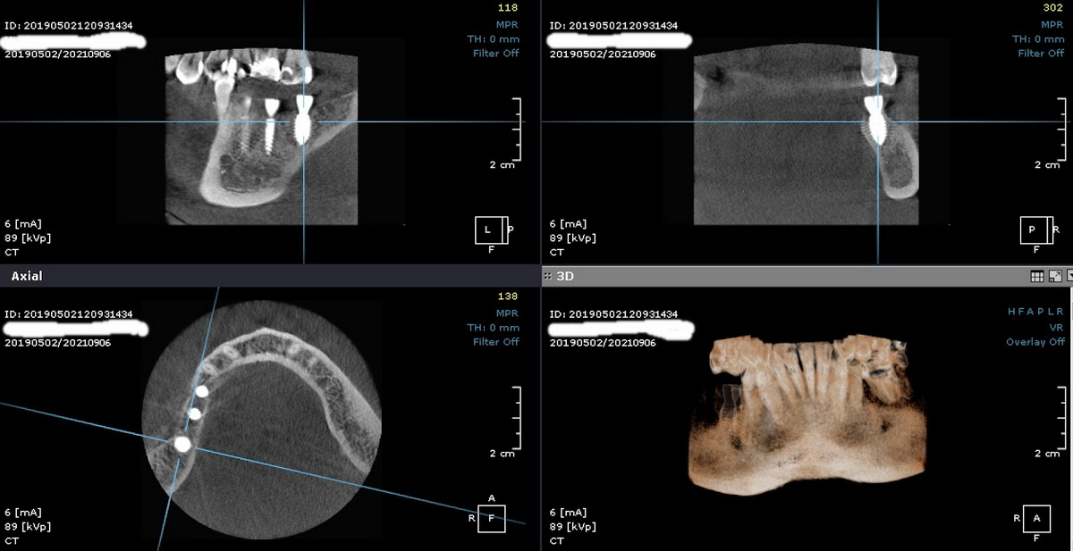
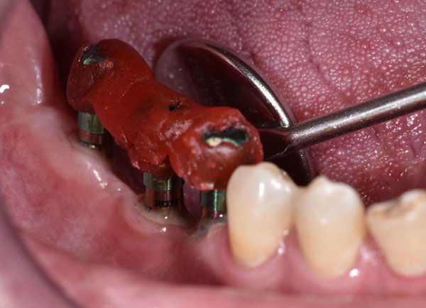
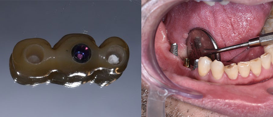
45-47 (FDI). It was decided to place two ROOTT R implants. 47d., the medullary part of the bone is soft ( D3 ) It was decided to use the R5510 implant with a special macro design, ideal for the circumstances mentioned above.
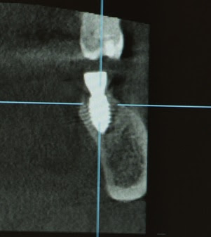
3D image before prosthesis, 3 months later.
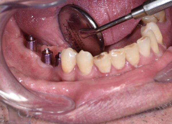
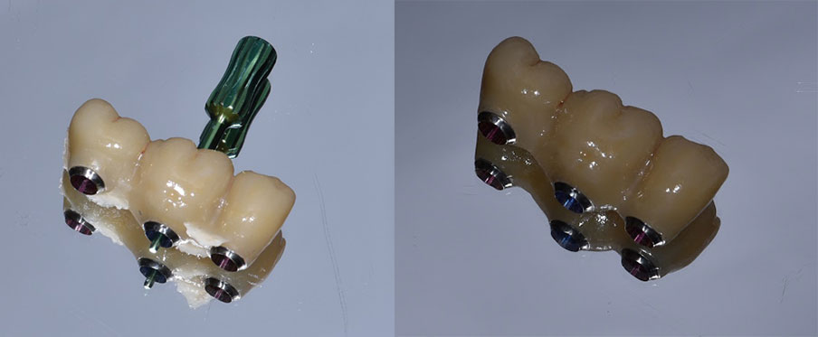
It was decided to use single-unit AK-type abutments because of the significant occlusal tooth height and the patient’s strong bite, which develops a high bite force.
The monoblock AK abutments were selected during the surgery and placed in the mouth with conometric mucosal shapers, according to the ONE ABUTMENT ONE TIME technique.
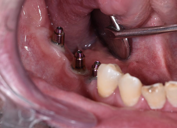
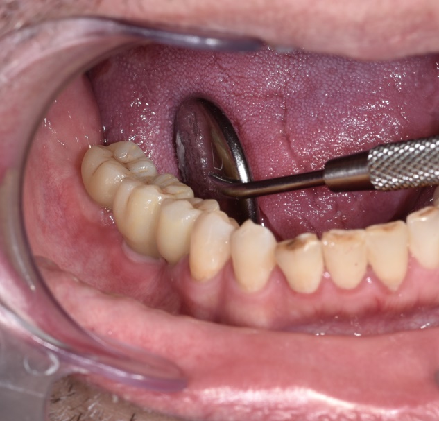
Pre-prosthetic view.
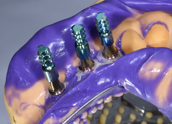
Observation of the formation of a properly fixed and keratinized mucosa around the implants
An analogue impression is taken with a composite material fixed around the conometric crowns, replicating the actual tissue profile around the implants
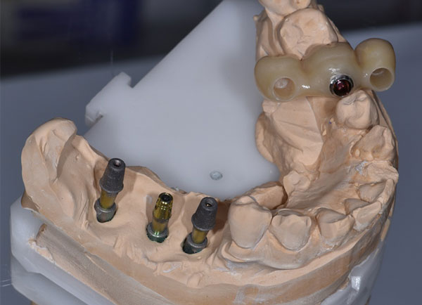
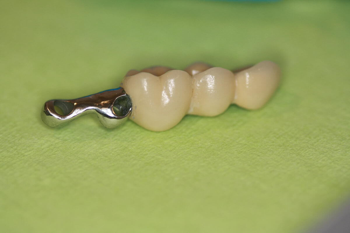
From a dental technician, zirconia bridge was received, in which one conometric cap was glued in. Another two will be secured in a clinical way.
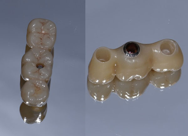
Conometric fixation of the bridge does not require screw fixation, so only one occlusal socket is observed instead of three.
Ready for cementation in the mouth. The cavities of the conometric crowns are sealed with Teflon. The closed shaft of the denture above 45 (FDI) tooth is also Teflon sealed.
After cementation in the mouth, the denture is removed with the help of a special instrument – the SR extractor – using the only open access hole located above 46 (FDI) tooth. After removing the excess cement, the prosthesis is ready for precise and passive fixation in the mouth.
The denture is fixed in the mouth. Can be fixed without the aid of screws. If desired, single screw fixation is possible in 46 (FDI) tooth, which was the only open access hole.